Since publication of Yasuda's and colleagues' [81] ret rospective study, many prospective studies comparing EUS to other imaging tests have shown that EUS is more accurate than any other test for diagnosis, staging, and predicting resectability of pancreatic tumors [78]. Most studies excluded patients with suspected bile duct stones and evaluated only patients suspected of having pancreatic disease. CT scan and standard US have a low sensitivity in finding bile duct stones. Including patients with bile duct stones into a study will naturally favor EUS because EUS has been shown to be very accurate in finding common bile duct stones [82]. Occasionally, the presentation of bile duct stones can mimic a tumor, but more than 80% can be detected using clinical and laboratory criteria alone [83]. Nevertheless, the greater accuracy of EUS to evaluate tumors, as compared with other imaging techniques, was also found in studies that did not include patients with common bile duct stones.
There are only 2 reports that studied the issue of clinical utility by prospectively comparing findings of EUS to the combined findings of CT plus ERCP to differentiate malignant from inflammatory pancreatobiliary tumors. One study [84] showed that EUS was superior, and the other study [85] showed that CT plus ERCP was better. In the study by Rosch and associates [85], mean tumor size was 4.5 cm (range, 2-20 cm). This study pointed out that, as reported by Yasuda and colleagues [81], EUS was superior to CT or ERCP for tumors measuring less than 3 cm. In the study by Snady and co-workers [84], in which EUS was superior to CT plus ERCP to differentiate malignant from inflammatory tumors, mean tumor size was less than 3 cm (maximum, 5 cm). Therefore, the primary difference between the 2 studies was the size of the lesion. Thus, in combining the data from both studies, it appears that the larger a tumor is, the less the difference between CT plus ERCP compared with EUS will be. For smaller, potentially resectable, tumors, the greater the difference will be in favor of EUS. Correctly identifying patients with resectable pancreatic carcinomas and no metastatic disease or vessel involvement would spare many others a major operation.
The size of a pancreatic tumor is the primary factor in determining an algorithm for diagnosis and treatment (Fig 10). Initial evaluation of suspected pancreatic carcinomas with ultrasound or CT scan will accurately stage the extent of disease for lesions larger than 5 cm. In pancreatobiliary disease, the accuracy of CT scan relies primarily on distortions of contiguous fat planes in the area. Difficulties with CT arise in evaluating small tumors. Fat planes may be unaltered because the tumor is still confined to the pancreas, or because there is an associated accompanying loss of fat planes with tumor-associated weight loss. Also, the density of small tumors is often so similar to that of the normal pancreatic parenchyma that it remains undetected despite thin-section CT scan. Although CT scan can visualize the pancreas in more than 90% of patients, a tumor must be at least 2 to 3 cm for probable detection. A mass may not even be detectable by CT scan in approximately 20% of patients preoperatively considered to have resectable carcinoma before EUS assessment.
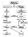
Figure 10
click to enlarge